What is inside a human eyeball
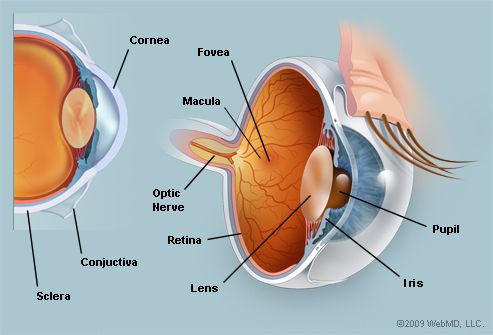
What Is Inside A Human Eyeball. The sclera is itself covered anteriorly by the conjunctiva a transparent mucous membrane that prevents the eye from. A clear watery fluid produced by the ciliary cody that fills the area between the lens and the cornea. The inside lining of the eye is covered by special light sensing cells that are collectively called the retina. Rod and cone cells in the retina are photoreceptive cells which are able to detect visible light and convey this information to the brain eyes signal information which is used by the brain to elicit the perception of color shape depth movement and other features.
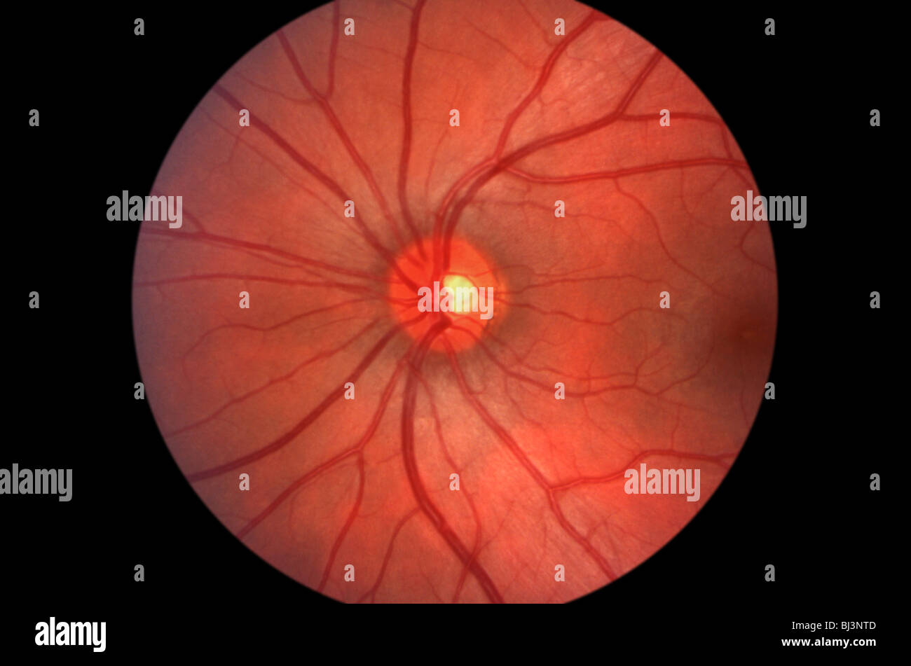 Medical Image Inside The Human Eye Showing The Retina Stock Photo Alamy From alamy.com
It converts light into electrical impulses. Rays of light enter the eye and an image of the object is focused on the retina. These allow the eye to move up and down and across while restricting movement so that the eye does not rotate back into the socket. The inside lining of the eye is covered by special light sensing cells that are collectively called the retina. The protrusion of the eyeballs proptosis in exophthalmic goitre is caused by the collection of fluid in the orbital fatty tissue. The internal structure of the eye.
The conjunctiva contains visible blood vessels that are visible against the white background of the sclera.
The internal structure of the eye. A clear watery fluid produced by the ciliary cody that fills the area between the lens and the cornea. The conjunctiva is a thin transparent layer of tissues covering the front of the eye including the sclera and the inside of the eyelids. The sclera the tough protective outer shell of the eyeball is composed of dense fibrous tissue that covers four fifths of the eyeball and provides attachments for the muscles that move the eye. The internal structure of the eye. The conjunctiva keeps bacteria and foreign material from getting behind the eye.
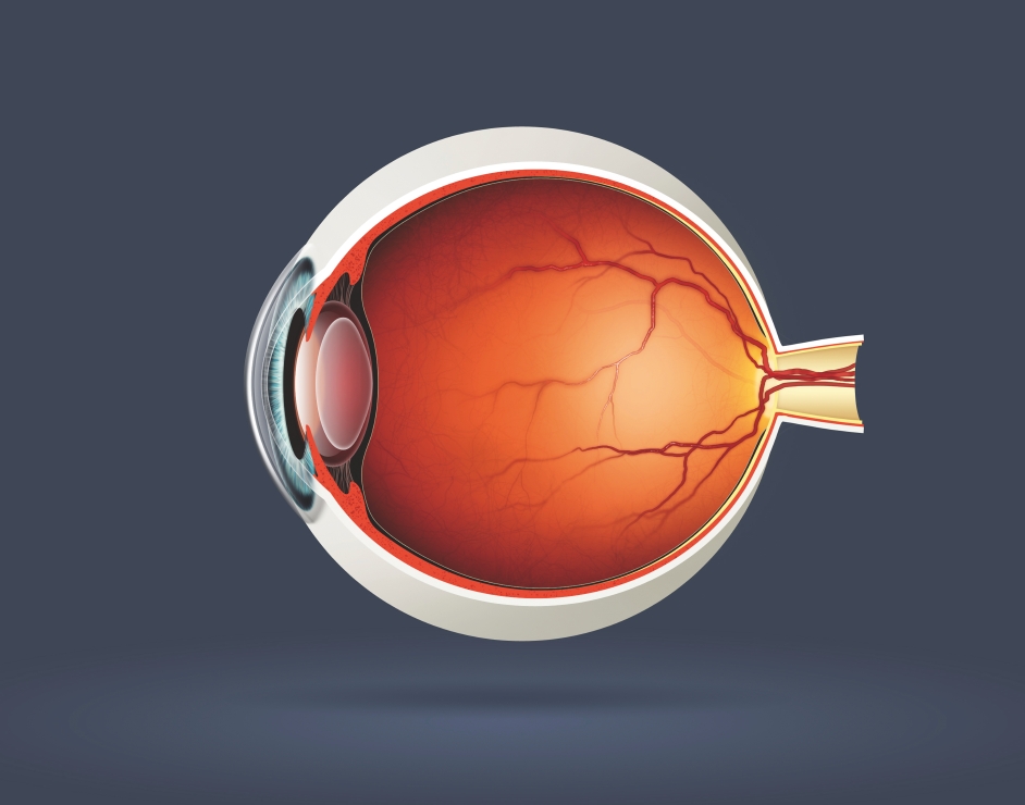 Source: howitworksdaily.com
The anterior segment is small accounting for some 20 of the inner area of the eyeball and lies between the cornea and anterior aspect front of the lens. The important structures within the eye includes the lens and suspensory ligaments and the aqeous and vitreous humor. The anterior segment is small accounting for some 20 of the inner area of the eyeball and lies between the cornea and anterior aspect front of the lens. The eyeball and its functional muscles are surrounded by a layer of orbital fat that acts much like a cushion permitting a smooth rotation of the eyeball about a virtually fixed point the centre of rotation. The conjunctiva contains visible blood vessels that are visible against the white background of the sclera.
 Source: alamy.com
The sclera the tough protective outer shell of the eyeball is composed of dense fibrous tissue that covers four fifths of the eyeball and provides attachments for the muscles that move the eye. The eye moves to allow a range of vision of approximately 180 degrees and to do this it has four primary muscles which control the movement of the eyeball. A clear watery fluid produced by the ciliary cody that fills the area between the lens and the cornea. The principle of light. These allow the eye to move up and down and across while restricting movement so that the eye does not rotate back into the socket.
 Source: shutterstock.com
This includes aqueous humour. The conjunctiva keeps bacteria and foreign material from getting behind the eye. These allow the eye to move up and down and across while restricting movement so that the eye does not rotate back into the socket. The principle of light. The human eye is a paired sense organ that reacts to light and allows vision.
 Source: nbcnews.com
The important structures within the eye includes the lens and suspensory ligaments and the aqeous and vitreous humor. The eyeball and its functional muscles are surrounded by a layer of orbital fat that acts much like a cushion permitting a smooth rotation of the eyeball about a virtually fixed point the centre of rotation. The eye moves to allow a range of vision of approximately 180 degrees and to do this it has four primary muscles which control the movement of the eyeball. The anterior segment is small accounting for some 20 of the inner area of the eyeball and lies between the cornea and anterior aspect front of the lens. These allow the eye to move up and down and across while restricting movement so that the eye does not rotate back into the socket.
 Source: webmd.com
Rays of light enter the eye and an image of the object is focused on the retina. This includes aqueous humour. The eye is part of the sensory nervous system. The important structures within the eye includes the lens and suspensory ligaments and the aqeous and vitreous humor. The protrusion of the eyeballs proptosis in exophthalmic goitre is caused by the collection of fluid in the orbital fatty tissue.
 Source: pinterest.com
The protrusion of the eyeballs proptosis in exophthalmic goitre is caused by the collection of fluid in the orbital fatty tissue. The retina inside human eye when a person sees an object either the object is giving off light or it is reflecting light from another source. The principle of light. The sclera is itself covered anteriorly by the conjunctiva a transparent mucous membrane that prevents the eye from. The human eye is a paired sense organ that reacts to light and allows vision.
 Source: quora.com
The inside lining of the eye is covered by special light sensing cells that are collectively called the retina. The principle of light. The conjunctiva keeps bacteria and foreign material from getting behind the eye. The internal structure of the eye. The anterior segment is small accounting for some 20 of the inner area of the eyeball and lies between the cornea and anterior aspect front of the lens.
 Source: arteinstitute.org
The important structures within the eye includes the lens and suspensory ligaments and the aqeous and vitreous humor. The eye is part of the sensory nervous system. The eye moves to allow a range of vision of approximately 180 degrees and to do this it has four primary muscles which control the movement of the eyeball. The internal structure of the eye. The sclera is itself covered anteriorly by the conjunctiva a transparent mucous membrane that prevents the eye from.
 Source: popularmechanics.com
There are two segments within the eyeball anterior segment and posterior segment. There are two segments within the eyeball anterior segment and posterior segment. The principle of light. A clear watery fluid produced by the ciliary cody that fills the area between the lens and the cornea. Rod and cone cells in the retina are photoreceptive cells which are able to detect visible light and convey this information to the brain eyes signal information which is used by the brain to elicit the perception of color shape depth movement and other features.
 Source: reddit.com
Rod and cone cells in the retina are photoreceptive cells which are able to detect visible light and convey this information to the brain eyes signal information which is used by the brain to elicit the perception of color shape depth movement and other features. The conjunctiva contains visible blood vessels that are visible against the white background of the sclera. It converts light into electrical impulses. The internal structure of the eye. A clear watery fluid produced by the ciliary cody that fills the area between the lens and the cornea.
 Source: youtube.com
The human eye is a paired sense organ that reacts to light and allows vision. The sclera is itself covered anteriorly by the conjunctiva a transparent mucous membrane that prevents the eye from. A clear watery fluid produced by the ciliary cody that fills the area between the lens and the cornea. Behind the eye your optic nerve carries. It converts light into electrical impulses.
 Source: en.wikipedia.org
The sclera is itself covered anteriorly by the conjunctiva a transparent mucous membrane that prevents the eye from. Behind the eye your optic nerve carries. The inside lining of the eye is covered by special light sensing cells that are collectively called the retina. The anterior segment is small accounting for some 20 of the inner area of the eyeball and lies between the cornea and anterior aspect front of the lens. The eyeball and its functional muscles are surrounded by a layer of orbital fat that acts much like a cushion permitting a smooth rotation of the eyeball about a virtually fixed point the centre of rotation.
 Source: pinterest.com
The eye is part of the sensory nervous system. The human eye is a paired sense organ that reacts to light and allows vision. A clear watery fluid produced by the ciliary cody that fills the area between the lens and the cornea. The eye is part of the sensory nervous system. The internal structure of the eye.
 Source: oconnortomsblog.wordpress.com
The sclera is itself covered anteriorly by the conjunctiva a transparent mucous membrane that prevents the eye from. Rays of light enter the eye and an image of the object is focused on the retina. Behind the eye your optic nerve carries. The inside lining of the eye is covered by special light sensing cells that are collectively called the retina. The eye is part of the sensory nervous system.
 Source: kidshealth.org
The important structures within the eye includes the lens and suspensory ligaments and the aqeous and vitreous humor. These allow the eye to move up and down and across while restricting movement so that the eye does not rotate back into the socket. A clear watery fluid produced by the ciliary cody that fills the area between the lens and the cornea. The protrusion of the eyeballs proptosis in exophthalmic goitre is caused by the collection of fluid in the orbital fatty tissue. This includes aqueous humour.
If you find this site adventageous, please support us by sharing this posts to your own social media accounts like Facebook, Instagram and so on or you can also bookmark this blog page with the title what is inside a human eyeball by using Ctrl + D for devices a laptop with a Windows operating system or Command + D for laptops with an Apple operating system. If you use a smartphone, you can also use the drawer menu of the browser you are using. Whether it’s a Windows, Mac, iOS or Android operating system, you will still be able to bookmark this website.


