Labelled cheek cell
Labelled Cheek Cell. Junior karimatsenga the human cheek cell 1. A cheek cell an epithelial cell found in the tissue on the inside lining of the mouth continually secretes mucus to maintains a moist environment in the mouth. Some of the worksheets for this concept are grade7lifescience lessonunitplanname microscopelab cellsmicroorganisms unit 3 weeks cell structure exploration activities chapter 12 bacteria protists and fungi plant and animal cells lab 3 use of the microscope introduction to biology lab. It is surrounded by cytoplasm.
 Ncert Class 9 Science Lab Manual Slide Of Onion Peel And Cheek Cells Cbse Tuts From cbsetuts.com
Go to google and type cheek cells into the search box. Junior karimatsenga the human cheek cell 1. Click on images to see all the. Sketch the cheek cell at medium and high power. Cheek cell labeled golfclub interphase cell cycle diagram vmglobalco labeled onion cell diagram wiring online 400x cheek cells 400x bacterial cells onioncells 400x elodea cells cheek cells under the microscope human cell drawing at getdrawingscom free for personal use human virtual plant animal cell lab mrs thomas classes. Sketch the image at scanning low and high power.
The nucleus at the central part of the cheek cell contains dna.
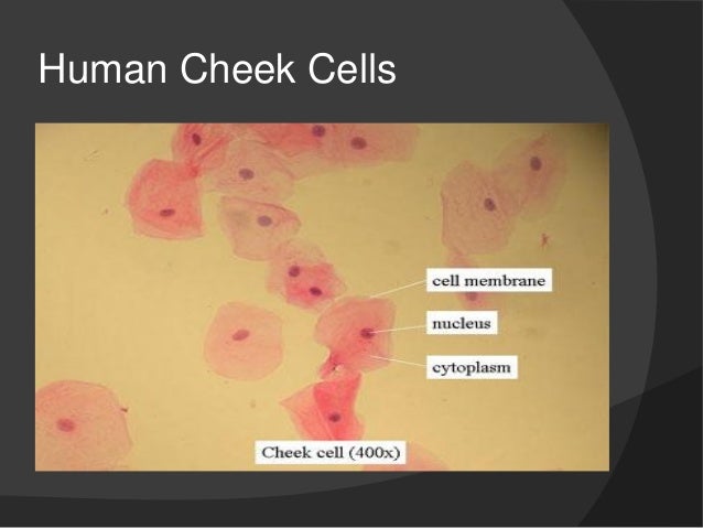
Identify and label the parts of human cheek cells displaying top 8 worksheets found for this concept. When a drop of methylene blue is introduced the nucleus is stained which makes it stand out and be clearly seen under the microscope. Label the nucleus cytoplasm and cell membrane. If you missed the microscope lab we did in class you will need to make it up by using a virtual microscope which can be accessed on the internet. Virtual microscope lab cheek cells introduction. Click on images to see all the.
 Source: studyrankers.com
The presence of a cell wall and a large vacuole are indicators that help identify plant cells such as seen in the onion peel. As in all animal cells the cells of the human cheek do not possess a cell wall. Some of the worksheets for this concept are grade7lifescience lessonunitplanname microscopelab cellsmicroorganisms unit 3 weeks cell structure exploration activities chapter 12 bacteria protists and fungi plant and animal cells lab 3 use of the microscope introduction to biology lab. In class we obtained cheek cells by scraping the inside of the mouth with a toothpick and then rubbing the toothpick on a drop of water with blue. Cheek cell labeled golfclub interphase cell cycle diagram vmglobalco labeled onion cell diagram wiring online 400x cheek cells 400x bacterial cells onioncells 400x elodea cells cheek cells under the microscope human cell drawing at getdrawingscom free for personal use human virtual plant animal cell lab mrs thomas classes.
 Source: slideshare.net
Identify and label the parts of human cheek cells displaying top 8 worksheets found for this concept. Virtual microscope lab cheek cells introduction. Sketch the image at scanning low and high power. Methylene blue stains negatively charged molecules in the cell including dna and rna. Label on high power the cell membrane cytoplasm and nucleus.
Source: hrewiringbrain.varosrl.it
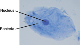
The cells seen are squamous epithelial cells from the outer epithelial layer of the mouth. Junior karimatsenga the human cheek cell 1. List the three main parts of the cell theory. The small blue dots are bacteria from our teeth and mouth. Click on images to see all the.
Source:
A cell membrane that is semi permeable surrounds the cytoplasm. Label the nucleus cytoplasm and cell membrane. In class we obtained cheek cells by scraping the inside of the mouth with a toothpick and then rubbing the toothpick on a drop of water with blue. A cell membrane that is semi permeable surrounds the cytoplasm. If you missed the microscope lab we did in class you will need to make it up by using a virtual microscope which can be accessed on the internet.
 Source: www2.mrc-lmb.cam.ac.uk
A cell membrane that is semi permeable surrounds the cytoplasm. Methylene blue stains negatively charged molecules in the cell including dna and rna. When a drop of methylene blue is introduced the nucleus is stained which makes it stand out and be clearly seen under the microscope. List the three main parts of the cell theory. All living things are composed of cells cells are the basic units of structure and function for living things all cells come from pre existing cells.
 Source: slideplayer.com
Virtual microscope lab cheek cells introduction. Cheek cell labeled golfclub interphase cell cycle diagram vmglobalco labeled onion cell diagram wiring online 400x cheek cells 400x bacterial cells onioncells 400x elodea cells cheek cells under the microscope human cell drawing at getdrawingscom free for personal use human virtual plant animal cell lab mrs thomas classes. Cells view the slide labeled cheek smear. Methylene blue stains negatively charged molecules in the cell including dna and rna. Some of the worksheets for this concept are grade7lifescience lessonunitplanname microscopelab cellsmicroorganisms unit 3 weeks cell structure exploration activities chapter 12 bacteria protists and fungi plant and animal cells lab 3 use of the microscope introduction to biology lab.
 Source: jacusers.johnabbott.qc.ca
The presence of a cell wall and a large vacuole are indicators that help identify plant cells such as seen in the onion peel. This dye is toxic when ingested and it causes irritation when in contact with the skin and eyes. Scanning 4 low 10 high 40 3. Junior karimatsenga the human cheek cell 1. Sketch the image at scanning low and high power.
 Source: cbsetuts.com
The nucleus at the central part of the cheek cell contains dna. A cell membrane that is semi permeable surrounds the cytoplasm. Click on images to see all the. Sketch the image at scanning low and high power. Sketch the cheek cell at medium and high power.
 Source: researchgate.net
If you missed the microscope lab we did in class you will need to make it up by using a virtual microscope which can be accessed on the internet. As in all animal cells the cells of the human cheek do not possess a cell wall. Junior karimatsenga the human cheek cell 1. Although the entire cell appears light blue in color the nucleus at the central part of the cell is much darker which allows it to be identified. In class we obtained cheek cells by scraping the inside of the mouth with a toothpick and then rubbing the toothpick on a drop of water with blue.
 Source: brainly.in
The nucleus at the central part of the cheek cell contains dna. Some of the worksheets for this concept are grade7lifescience lessonunitplanname microscopelab cellsmicroorganisms unit 3 weeks cell structure exploration activities chapter 12 bacteria protists and fungi plant and animal cells lab 3 use of the microscope introduction to biology lab. When a drop of methylene blue is introduced the nucleus is stained which makes it stand out and be clearly seen under the microscope. Label on high power the cell membrane cytoplasm and nucleus. It is surrounded by cytoplasm.
 Source: microcosmos.foldscope.com
The cells seen are squamous epithelial cells from the outer epithelial layer of the mouth. If you missed the microscope lab we did in class you will need to make it up by using a virtual microscope which can be accessed on the internet. As in all animal cells the cells of the human cheek do not possess a cell wall. Scanning 4 low 10 high 40 3. The small blue dots are bacteria from our teeth and mouth.
 Source: brainly.in
Click on images to see all the. This dye is toxic when ingested and it causes irritation when in contact with the skin and eyes. A cell membrane that is semi permeable surrounds the cytoplasm. It is surrounded by cytoplasm. All living things are composed of cells cells are the basic units of structure and function for living things all cells come from pre existing cells.
 Source: fankhauserblog.wordpress.com
It is surrounded by cytoplasm. The cells seen are squamous epithelial cells from the outer epithelial layer of the mouth. This dye is toxic when ingested and it causes irritation when in contact with the skin and eyes. Cells view the slide labeled cheek smear. Together with salivary glands that secrete saliva the cheek cells supply enough moisture in the mouth for enzymes to thrive.
 Source: schoolworkhelper.net
As in all animal cells the cells of the human cheek do not possess a cell wall. Cheek cell labeled golfclub interphase cell cycle diagram vmglobalco labeled onion cell diagram wiring online 400x cheek cells 400x bacterial cells onioncells 400x elodea cells cheek cells under the microscope human cell drawing at getdrawingscom free for personal use human virtual plant animal cell lab mrs thomas classes. Although the entire cell appears light blue in color the nucleus at the central part of the cell is much darker which allows it to be identified. It is surrounded by cytoplasm. List the three main parts of the cell theory.
 Source: youtube.com
Junior karimatsenga the human cheek cell 1. The presence of a cell wall and a large vacuole are indicators that help identify plant cells such as seen in the onion peel. This dye is toxic when ingested and it causes irritation when in contact with the skin and eyes. All living things are composed of cells cells are the basic units of structure and function for living things all cells come from pre existing cells. Label the nucleus cytoplasm and cell membrane.
If you find this site good, please support us by sharing this posts to your preference social media accounts like Facebook, Instagram and so on or you can also bookmark this blog page with the title labelled cheek cell by using Ctrl + D for devices a laptop with a Windows operating system or Command + D for laptops with an Apple operating system. If you use a smartphone, you can also use the drawer menu of the browser you are using. Whether it’s a Windows, Mac, iOS or Android operating system, you will still be able to bookmark this website.


