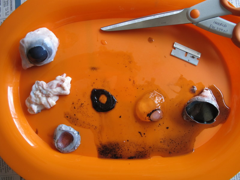
Human eyeball dissection
Human Eyeball Dissection. These accessory organs include the eyelids and lacrimal apparatus which protect the eye and a set of extrinsic muscles which move the eye. The anatomy of the eye is fascinating and this quiz game will help you memorize the 12 parts of the eye with ease. The anatomy of the human eye can be better shown and understood by the actual dissection of an eye. Light enters our eyes through the pupil then passes through a lens and the fluid filled vitreous.
 Cow Eye Dissection Student Cut 2 For Lesson Plan Youtube From youtube.com
The eye science quiz. The human eye the organ containing visual receptors provides vision with the assistance of accessory organs. Vision is by far the most used of the five senses and is one of the primary means that we use to gather information from our surroundings. More than 75. Light enters our eyes through the pupil then passes through a lens and the fluid filled vitreous. Sheep eyes are removed at the time the animal is slaughtered and then preserved for later use.
The eye science quiz.
The eye is part of the sensory nervous system. How do your eyes work. Sheep eyes are removed at the time the animal is slaughtered and then preserved for later use. More than 75. Join ross exton on a journey looking inside a horse eyeball investigating the anatomy of this fascinating organ along the way this vi. The eyeball and its functional muscles are surrounded by a layer of orbital fat that acts much like a cushion permitting a smooth rotation of the eyeball about a virtually fixed point the centre of rotation.
 Source: ingridscience.ca
The anatomy of the human eye can be better shown and understood by the actual dissection of an eye. The human eye the organ containing visual receptors provides vision with the assistance of accessory organs. The eye science quiz. The anatomy of the human eye can be better shown and understood by the actual dissection of an eye. The eyeball and its functional muscles are surrounded by a layer of orbital fat that acts much like a cushion permitting a smooth rotation of the eyeball about a virtually fixed point the centre of rotation.
 Source: homesciencetools.com
Procedural video showing how to dissect the eye and identify the major structures such as the lens iris pupil vitreous humor retina and tapetum. The eyeball and its functional muscles are surrounded by a layer of orbital fat that acts much like a cushion permitting a smooth rotation of the eyeball about a virtually fixed point the centre of rotation. These accessory organs include the eyelids and lacrimal apparatus which protect the eye and a set of extrinsic muscles which move the eye. Rod and cone cells in the retina are photoreceptive cells which are able to detect visible light and convey this information to the brain eyes signal information which is used by the brain to elicit the perception of color shape depth movement and other features. More than 75.

Rod and cone cells in the retina are photoreceptive cells which are able to detect visible light and convey this information to the brain eyes signal information which is used by the brain to elicit the perception of color shape depth movement and other features. The eyeball and its functional muscles are surrounded by a layer of orbital fat that acts much like a cushion permitting a smooth rotation of the eyeball about a virtually fixed point the centre of rotation. Parts of the eye. Rod and cone cells in the retina are photoreceptive cells which are able to detect visible light and convey this information to the brain eyes signal information which is used by the brain to elicit the perception of color shape depth movement and other features. Although the eye is small only about 1 inch in diameter each part plays an important role in allowing people to see the world.

Light enters our eyes through the pupil then passes through a lens and the fluid filled vitreous. Dissection of sheep eyeball. The eye science quiz. Rod and cone cells in the retina are photoreceptive cells which are able to detect visible light and convey this information to the brain eyes signal information which is used by the brain to elicit the perception of color shape depth movement and other features. One eye of choice for dissection that closely resembles the human eye is that of the sheep.
 Source: exploratorium.edu
Sheep eyes are removed at the time the animal is slaughtered and then preserved for later use. Light enters our eyes through the pupil then passes through a lens and the fluid filled vitreous. The human eye the organ containing visual receptors provides vision with the assistance of accessory organs. The eye is part of the sensory nervous system. Parts of the eye.

The eye is part of the sensory nervous system. Procedural video showing how to dissect the eye and identify the major structures such as the lens iris pupil vitreous humor retina and tapetum. These accessory organs include the eyelids and lacrimal apparatus which protect the eye and a set of extrinsic muscles which move the eye. Dissection of sheep eyeball. The anatomy of the eye is fascinating and this quiz game will help you memorize the 12 parts of the eye with ease.
 Source: benandme.com
How do your eyes work. Eye anatomy function and physiology facts. The anatomy of the eye is fascinating and this quiz game will help you memorize the 12 parts of the eye with ease. Dissection of sheep eyeball. The human eye the organ containing visual receptors provides vision with the assistance of accessory organs.
 Source: youtube.com
One eye of choice for dissection that closely resembles the human eye is that of the sheep. Procedural video showing how to dissect the eye and identify the major structures such as the lens iris pupil vitreous humor retina and tapetum. Dissection of sheep eyeball. The eye is part of the sensory nervous system. These accessory organs include the eyelids and lacrimal apparatus which protect the eye and a set of extrinsic muscles which move the eye.
 Source: youtube.com
These accessory organs include the eyelids and lacrimal apparatus which protect the eye and a set of extrinsic muscles which move the eye. Rod and cone cells in the retina are photoreceptive cells which are able to detect visible light and convey this information to the brain eyes signal information which is used by the brain to elicit the perception of color shape depth movement and other features. The human eye the organ containing visual receptors provides vision with the assistance of accessory organs. More than 75. The protrusion of the eyeballs proptosis in exophthalmic goitre is caused by the collection of fluid in the orbital fatty tissue.
 Source: science.jburroughs.org
Sheep eyes are removed at the time the animal is slaughtered and then preserved for later use. The human eye is a paired sense organ that reacts to light and allows vision. The anatomy of the human eye can be better shown and understood by the actual dissection of an eye. How do your eyes work. The human eye the organ containing visual receptors provides vision with the assistance of accessory organs.
 Source: exploratorium.edu
One eye of choice for dissection that closely resembles the human eye is that of the sheep. The eyeball and its functional muscles are surrounded by a layer of orbital fat that acts much like a cushion permitting a smooth rotation of the eyeball about a virtually fixed point the centre of rotation. Light enters our eyes through the pupil then passes through a lens and the fluid filled vitreous. More than 75. Eye anatomy function and physiology facts.
 Source: youtube.com
Procedural video showing how to dissect the eye and identify the major structures such as the lens iris pupil vitreous humor retina and tapetum. One eye of choice for dissection that closely resembles the human eye is that of the sheep. Although the eye is small only about 1 inch in diameter each part plays an important role in allowing people to see the world. Join ross exton on a journey looking inside a horse eyeball investigating the anatomy of this fascinating organ along the way this vi. How do your eyes work.
 Source: exploratorium.edu
Our eyes are highly specialized organs that take in the light reflected off our surroundings and transform it into electrical impulses to send to the brain. Vision is by far the most used of the five senses and is one of the primary means that we use to gather information from our surroundings. Our eyes are highly specialized organs that take in the light reflected off our surroundings and transform it into electrical impulses to send to the brain. Procedural video showing how to dissect the eye and identify the major structures such as the lens iris pupil vitreous humor retina and tapetum. Eye anatomy function and physiology facts.
 Source: quizlet.com
Join ross exton on a journey looking inside a horse eyeball investigating the anatomy of this fascinating organ along the way this vi. One eye of choice for dissection that closely resembles the human eye is that of the sheep. Rod and cone cells in the retina are photoreceptive cells which are able to detect visible light and convey this information to the brain eyes signal information which is used by the brain to elicit the perception of color shape depth movement and other features. Our eyes are highly specialized organs that take in the light reflected off our surroundings and transform it into electrical impulses to send to the brain. The eye is part of the sensory nervous system.
 Source: smithsonianmag.com
Eye anatomy function and physiology facts. Dissection of sheep eyeball. Eye anatomy function and physiology facts. The anatomy of the human eye can be better shown and understood by the actual dissection of an eye. Rod and cone cells in the retina are photoreceptive cells which are able to detect visible light and convey this information to the brain eyes signal information which is used by the brain to elicit the perception of color shape depth movement and other features.
If you find this site adventageous, please support us by sharing this posts to your own social media accounts like Facebook, Instagram and so on or you can also bookmark this blog page with the title human eyeball dissection by using Ctrl + D for devices a laptop with a Windows operating system or Command + D for laptops with an Apple operating system. If you use a smartphone, you can also use the drawer menu of the browser you are using. Whether it’s a Windows, Mac, iOS or Android operating system, you will still be able to bookmark this website.


