Electron microscope invented
Electron Microscope Invented. The electron microscope was invented in the early 1930s to overcome the limitations of light microscopes. By directing electrons at an object and measuring the frequency at which they bounce back an image can be formed. An image is formed from the interaction of the electrons with the sample as the beam is transmitted through the specimen. Microscopy is the science of.
 What Is An Electron Microscope What Does It Do From polymersolutions.com
Using magnetic lenses that direct electrons these electron waves were ultimately captured and then directed at objects. μικρός mikrós small and σκοπεῖν skopeîn to look or see is a laboratory instrument used to examine objects that are too small to be seen by the naked eye. The other half of the nobel prize was divided between heinrich rohrer and gerd binnig for the stm. The electron microscope. Microscopy is the science of. Over the course of nearly two decades three researchers jacques dubochet joachim franck and richard henderson created a technique for generating a 3d structure of the protein at an atomic level using an electron microscope.
Although max knoll produced a photo with a 50 mm object field width showing channeling contrast by the use of an electron beam scanner it was manfred von ardenne who in 1937 invented a microscope with high resolution by scanning a very small raster with a demagnified and finely focused.
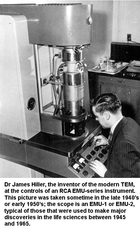
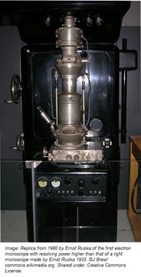
Siemens schuckertwerke released the first commercial electron microscope to the public in 1938. An account of the early history of scanning electron microscopy has been presented by mcmullan. The first north american electron microscope was constructed in 1938 at the university of toronto by eli franklin burton and students cecil hall james hillier and albert prebus. Microscope microscope uses small sample observation notable experiments discovery of cells related items optical microscope electron microscope a microscope from the ancient greek. This discovery initiated the study of electron optics and by 1931 german electrical engineers max knoll and ernst ruska had devised a two lens electron microscope that produced images of the electron source. This led to the first electron microscope.
 Source: papertrell.com
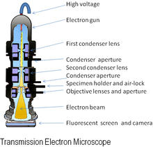
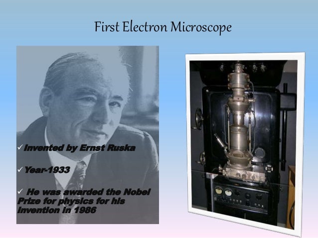
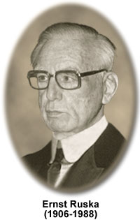
Over the course of nearly two decades three researchers jacques dubochet joachim franck and richard henderson created a technique for generating a 3d structure of the protein at an atomic level using an electron microscope. Siemens produced a transmission electron microscope tem in 1939. The introduction of the electron microscope in the 1930 s filled the bill. Transmission electron microscopy tem is a microscopy technique in which a beam of electrons is transmitted through a specimen to form an image. Co invented by germans max knoll and ernst ruska in 1931 ernst ruska was awarded half of the nobel prize for physics in 1986 for his invention.
 Source: ernstruska.de
An account of the early history of scanning electron microscopy has been presented by mcmullan. The electron microscope was invented in the early 1930s to overcome the limitations of light microscopes. Siemens schuckertwerke released the first commercial electron microscope to the public in 1938. Siemens produced the first commercial electron microscope in 1938. It was originally invented to view nonbiological materials such as metals.
 Source: en.wikipedia.org
This led to the first electron microscope. An account of the early history of scanning electron microscopy has been presented by mcmullan. The specimen is most often an ultrathin section less than 100 nm thick or a suspension on a grid. By directing electrons at an object and measuring the frequency at which they bounce back an image can be formed. The introduction of the electron microscope in the 1930 s filled the bill.
 Source: study.com
Although max knoll produced a photo with a 50 mm object field width showing channeling contrast by the use of an electron beam scanner it was manfred von ardenne who in 1937 invented a microscope with high resolution by scanning a very small raster with a demagnified and finely focused. In the same year manfred von ardenne developed the first scanning electron microscope. The first north american electron microscope was constructed in 1938 at the university of toronto by eli franklin burton and students cecil hall james hillier and albert prebus. It was originally invented to view nonbiological materials such as metals. Microscope microscope uses small sample observation notable experiments discovery of cells related items optical microscope electron microscope a microscope from the ancient greek.
 Source: sites.google.com
The introduction of the electron microscope in the 1930 s filled the bill. This discovery initiated the study of electron optics and by 1931 german electrical engineers max knoll and ernst ruska had devised a two lens electron microscope that produced images of the electron source. The specimen is most often an ultrathin section less than 100 nm thick or a suspension on a grid. Microscopy is the science of. Siemens produced a transmission electron microscope tem in 1939.
 Source: aaas.org
An image is formed from the interaction of the electrons with the sample as the beam is transmitted through the specimen. Siemens produced the first commercial electron microscope in 1938. The electron microscope. The electron microscope was invented in the early 1930s to overcome the limitations of light microscopes. By directing electrons at an object and measuring the frequency at which they bounce back an image can be formed.
 Source: doctorc.net
It was originally invented to view nonbiological materials such as metals. The introduction of the electron microscope in the 1930 s filled the bill. Although max knoll produced a photo with a 50 mm object field width showing channeling contrast by the use of an electron beam scanner it was manfred von ardenne who in 1937 invented a microscope with high resolution by scanning a very small raster with a demagnified and finely focused. Siemens produced a transmission electron microscope tem in 1939. The other half of the nobel prize was divided between heinrich rohrer and gerd binnig for the stm.
 Source: polymersolutions.com
Over the course of nearly two decades three researchers jacques dubochet joachim franck and richard henderson created a technique for generating a 3d structure of the protein at an atomic level using an electron microscope. The introduction of the electron microscope in the 1930 s filled the bill. Co invented by germans max knoll and ernst ruska in 1931 ernst ruska was awarded half of the nobel prize for physics in 1986 for his invention. The specimen is most often an ultrathin section less than 100 nm thick or a suspension on a grid. μικρός mikrós small and σκοπεῖν skopeîn to look or see is a laboratory instrument used to examine objects that are too small to be seen by the naked eye.
 Source: azom.com
Siemens schuckertwerke released the first commercial electron microscope to the public in 1938. An account of the early history of scanning electron microscopy has been presented by mcmullan. The other half of the nobel prize was divided between heinrich rohrer and gerd binnig for the stm. Siemens schuckertwerke released the first commercial electron microscope to the public in 1938. Light microscopes at that time could magnify specimens as high as 1000 times.
 Source: en.wikipedia.org
Transmission electron microscopy tem is a microscopy technique in which a beam of electrons is transmitted through a specimen to form an image. Siemens schuckertwerke released the first commercial electron microscope to the public in 1938. The electron microscope was invented in the early 1930s to overcome the limitations of light microscopes. Siemens produced a transmission electron microscope tem in 1939. Light microscopes at that time could magnify specimens as high as 1000 times.
 Source: news-medical.net
Transmission electron microscopy tem is a microscopy technique in which a beam of electrons is transmitted through a specimen to form an image. The electron microscope. This led to the first electron microscope. The first north american electron microscope was constructed in 1938 at the university of toronto by eli franklin burton and students cecil hall james hillier and albert prebus. Light microscopes at that time could magnify specimens as high as 1000 times.
 Source: slideshare.net
Co invented by germans max knoll and ernst ruska in 1931 ernst ruska was awarded half of the nobel prize for physics in 1986 for his invention. In the same year manfred von ardenne developed the first scanning electron microscope. It was originally invented to view nonbiological materials such as metals. Co invented by germans max knoll and ernst ruska in 1931 ernst ruska was awarded half of the nobel prize for physics in 1986 for his invention. By directing electrons at an object and measuring the frequency at which they bounce back an image can be formed.
 Source: leo-em.co.uk
An account of the early history of scanning electron microscopy has been presented by mcmullan. The electron microscope was invented in the early 1930s to overcome the limitations of light microscopes. Over the course of nearly two decades three researchers jacques dubochet joachim franck and richard henderson created a technique for generating a 3d structure of the protein at an atomic level using an electron microscope. Siemens produced a transmission electron microscope tem in 1939. This discovery initiated the study of electron optics and by 1931 german electrical engineers max knoll and ernst ruska had devised a two lens electron microscope that produced images of the electron source.
 Source: eurekalert.org
The specimen is most often an ultrathin section less than 100 nm thick or a suspension on a grid. The introduction of the electron microscope in the 1930 s filled the bill. Although max knoll produced a photo with a 50 mm object field width showing channeling contrast by the use of an electron beam scanner it was manfred von ardenne who in 1937 invented a microscope with high resolution by scanning a very small raster with a demagnified and finely focused. Siemens produced the first commercial electron microscope in 1938. Light microscopes at that time could magnify specimens as high as 1000 times.
 Source: en.wikipedia.org
In 1933 a primitive electron microscope was built that imaged a specimen rather than the electron source and in 1935 knoll produced a scanned image of a solid surface. An account of the early history of scanning electron microscopy has been presented by mcmullan. Siemens schuckertwerke released the first commercial electron microscope to the public in 1938. Light microscopes at that time could magnify specimens as high as 1000 times. This led to the first electron microscope.
If you find this site helpful, please support us by sharing this posts to your preference social media accounts like Facebook, Instagram and so on or you can also bookmark this blog page with the title electron microscope invented by using Ctrl + D for devices a laptop with a Windows operating system or Command + D for laptops with an Apple operating system. If you use a smartphone, you can also use the drawer menu of the browser you are using. Whether it’s a Windows, Mac, iOS or Android operating system, you will still be able to bookmark this website.


