Cow eye diagram
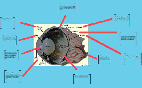
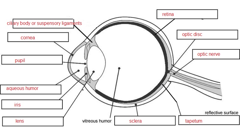
Cow Eye Diagram. A step by step hints and tips a cow eye primer and a glossary of terms. Tapetum mid layer of the eye and contains reflective pigments that helps the eye see better at night. A step by step hints and tips a cow eye primer and a glossary of terms. Start studying cow eye.
 Cow Eye Dissection Cow Eyes Eye Structure Eyeball Diagram From pinterest.com
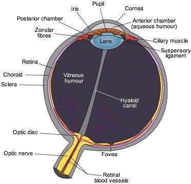
Takes electrical signals to the brain. It reflects light back through the retina. L ens clear flexible structure that focuses the light on the retina. Aug 30 2012 learn how to dissect a cow s eye in your classroom. Add to favorites 0 favs. The optic nerve s job is to send information from that was accumulated in the eye to the brain so you can understand what you are looking at.
About press copyright contact us creators advertise developers terms privacy policy safety how youtube works test new features press copyright contact us creators.
Online quiz to learn cow s eye diagram. The area around the fovea 4 retina. C horoid layer mid layer of the eye and contains dark pigments that helps nourish the retina. Online quiz to learn cow s eye diagram. Aug 30 2012 learn how to dissect a cow s eye in your classroom. You need to get 100 to score the 11 points available.
 Source: prezi.com
Add to favorites 0 favs. Notice that the retina s blood supply comes in through the center of the optic nerve. The back of the inside of the eyeball this is where the light sensitive rods and cones are located. The human eye 1 optic nerve. C horoid layer mid layer of the eye and contains dark pigments that helps nourish the retina.
 Source: pinterest.com
A step by step hints and tips a cow eye primer and a glossary of terms. The colorful and shiny part of the eye that lets the cow see in the dark and is located behind the retina. Your skills rank. Start studying cow eye. Although they do not show it here there is one more part of the eye called the optic nerve that lies on the anterior of the eye.
 Source: exploratorium.edu
Start studying cow eye. Start studying cow eye. The back of the inside of the eyeball this is where the light sensitive rods and cones are located. The optic nerve s job is to send information from that was accumulated in the eye to the brain so you can understand what you are looking at. The eyeball is the whole shell that keeps every part in place.
 Source: id.pinterest.com
Aug 30 2012 learn how to dissect a cow s eye in your classroom. L ens clear flexible structure that focuses the light on the retina. The area around the fovea 4 retina. Your skills rank. The eyeball is the whole shell that keeps every part in place.
 Source: exploratorium.edu
The sclera is the thick white outer covering of the eye. A step by step hints and tips a cow eye primer and a glossary of terms. L ens clear flexible structure that focuses the light on the retina. Aug 30 2012 learn how to dissect a cow s eye in your classroom. S clera a tough white outermost covering of the eye.
 Source: macroevolution.net
A step by step hints and tips a cow eye primer and a glossary of terms. Cow s eye diagram learn by taking a quiz. The eyeball is the whole shell that keeps every part in place. Notice that the retina s blood supply comes in through the center of the optic nerve. C horoid layer mid layer of the eye and contains dark pigments that helps nourish the retina.
 Source: quizlet.com
Your skills rank. The sclera is the thick white outer covering of the eye. Learn how to dissect a cow s eye in your classroom. A step by step hints and tips a cow eye primer and a glossary of terms. Your skills rank.
 Source: pinterest.com
A step by step hints and tips a cow eye primer and a glossary of terms. Although they do not show it here there is one more part of the eye called the optic nerve that lies on the anterior of the eye. Your skills rank. Learn how to dissect a cow s eye in your classroom. A step by step hints and tips a cow eye primer and a glossary of terms.
 Source: dissectingacoweye.weebly.com
Aug 30 2012 learn how to dissect a cow s eye in your classroom. Learn how to dissect a cow s eye in your classroom. Notice that the retina s blood supply comes in through the center of the optic nerve. S clera a tough white outermost covering of the eye. A step by step hints and tips a cow eye primer and a glossary of terms.
 Source: kata-kata-pujian.blogspot.com
Aug 30 2012 learn how to dissect a cow s eye in your classroom. The optic nerve s job is to send information from that was accumulated in the eye to the brain so you can understand what you are looking at. The area around the fovea 4 retina. Start studying cow eye. The back of the inside of the eyeball this is where the light sensitive rods and cones are located.
 Source: biologycorner.com
A step by step hints and tips a cow eye primer and a glossary of terms. You need to get 100 to score the 11 points available. Online quiz to learn cow s eye diagram. Learn how to dissect a cow s eye in your classroom. Start studying cow eye.
Source: docs.google.com
Tapetum mid layer of the eye and contains reflective pigments that helps the eye see better at night. The sclera is the thick white outer covering of the eye. Takes electrical signals to the brain. Learn vocabulary terms and more with flashcards games and other study tools. About press copyright contact us creators advertise developers terms privacy policy safety how youtube works test new features press copyright contact us creators.
 Source: pinterest.com
Focal point the center of your vision 3 macula. Learn how to dissect a cow s eye in your classroom. It reflects light back through the retina. Aug 30 2012 learn how to dissect a cow s eye in your classroom. The eyeball is the whole shell that keeps every part in place.
 Source: exploratorium.edu
Start studying cow eye. Your skills rank. Notice that the retina s blood supply comes in through the center of the optic nerve. It reflects light back through the retina. The colorful and shiny part of the eye that lets the cow see in the dark and is located behind the retina.
 Source: slideshare.net
The back of the inside of the eyeball this is where the light sensitive rods and cones are located. It reflects light back through the retina. Learn how to dissect a cow s eye in your classroom. You need to get 100 to score the 11 points available. Learn how to dissect a cow s eye in your classroom.
If you find this site good, please support us by sharing this posts to your preference social media accounts like Facebook, Instagram and so on or you can also save this blog page with the title cow eye diagram by using Ctrl + D for devices a laptop with a Windows operating system or Command + D for laptops with an Apple operating system. If you use a smartphone, you can also use the drawer menu of the browser you are using. Whether it’s a Windows, Mac, iOS or Android operating system, you will still be able to bookmark this website.


